Türkçe
 English
 German
 Spanish

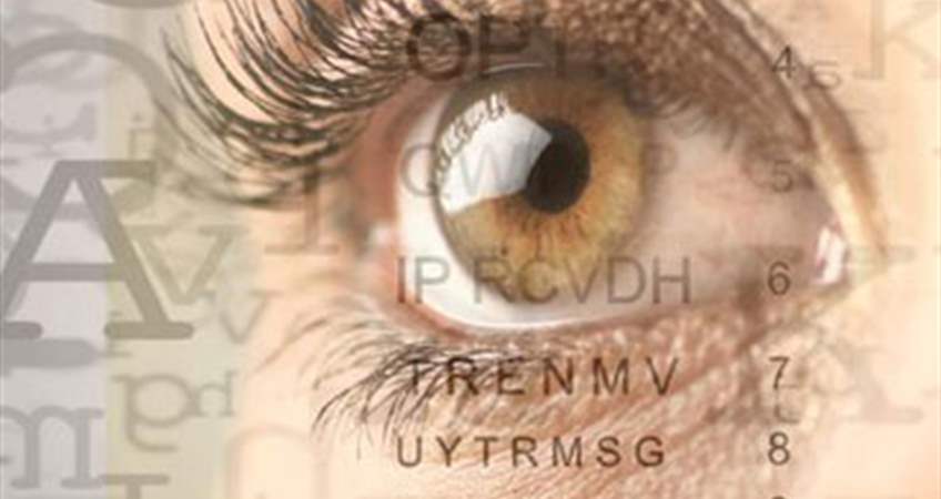
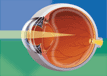
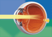
Normal Eye
80% of the light reflecting an object is refracted through the cornea (transport layer) layer of the eye, and 20% is refracted through the eye lens and focuses on a point over retina (nerve net layer). The stimulations the retina are transmitted to the eye nerve and brain forming image. This travel of the refracted light in the eye is called refraction. If the light is not focused on the right place in the retina, refractive errors are developed such as myopia, hypermetropia and astigmatism, and the clarity of sight is lost.
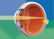
Myopia
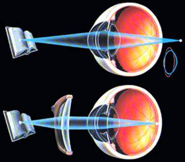
 If the anteroposterior length and cornea refractivity of the eye or both of them are more than they should be, the images of the objects are created in front of the macular area. Although myopic people are near-sighted, they have a blurred foresight.
The reasons:
1- Genetic; If the mother or father has myopia, the possibility for the children to develop myopia is increased.
2- Stress; Myopia may observed visual stress, i.e. in people who are engaged in reading, intensive computer works and close-range businesses.
3- In company with disease; Myopia may be developed adventitiously in people with diabetes mellitus or cataract.
4- Pseudomyopia; It is the case in which myopia development takes place in the eyes due to that ciliary muscles in the eye are not completely relaxed as a result of stress, fatigue and overload. It may sometimes take 2-3 days or sometimes it may last for years. Therefore, it is helpful and significant to enlarge eye pupils and temporarily paralyze ciliary muscles in to differentiate real myopia.
Myopia Disease - Malignant Myopia
In respect of some myopic patients with huge eye numbers, the problem is not only composed of the diopter value. Normal myopia is generally up to 7 degrees; it stops at about the age of 18-20; visual acuity is good and retina is normal. Malignant Myopia progresses regardless of age and the number may be high as 10-30. Also, the vision may remain at low levels in those patients due to the pathological changes in the retinal layer, retinal attenuation and destruction in the eye nerve and macula (see. Retina Diseases).
Hypermetropia
 If the anteroposterior length and corneal curvature, i.e. refractivity, or both of them are less than they should be, the images are created behind the macular area. The lens within the eye changes shape (accommodation) increasing its refractivity and tries to bring the image on the macular area. If hypermetropia could be compensated by the lens the person may have both near- and foresight, or have a foresight while having a blurred nearsight. At the same time, as the small muscles work too much to ensure that the lens changes shape, an early fatigue feeling, eye pain and headache may develop in the eye. The ability of the lens to change shape is reduced as the age advances despite the fact that it is high during childhood and adolescence.
The reasons:
It is generally genetic. If it is not realized at early ages, it may result in amblyopia or strabismus.
Astigmatism
 Astigmatism is mostly due to corneal deformity which is basically the egg shape of the cornea. This situation leads to different refractions in two axes; for example, if the number of an axis is 1 while the other one is 3, the difference between them is the astigmatism result and the value of it is 3-1=2. Astigmatism results in blurred or double vision. Cylindrical glasses are used for its treatment.
The reasons:
Genetic
Chalazion, deformity
Pterygium; the growth of vascular membrane tissue in the eye called conjunctiva overflowing the cornea.
Scars due to corneal infections
Keratoconus Disease
Presbyopia
It is mistaken for hypermetropia but this situation is the case of farsightedness due to the aging of the lens which also appears in people without visual impairment. The lens has to have a particular flexibility to change form for being able to focus on close-range. Due to aging, lens loses its flexibility. Presbyopia starts at the age of 40-45. This situation develops at an earlier age in people with hypermetropia. Myopic people realize that they are able to read better when they take their glasses off.
 Age-Related Visual Impairment = Presbyopia
It is mistaken for hypermetropia but this situation is the case of farsightedness due to the aging of the lens which also appears in people without visual impairment. The lens has to have a particular flexibility to change form for being able to focus on close-range. Due to aging, lens loses its flexibility. Presbyopia starts at the age of 40-45. This situation develops at an earlier age in people with hypermetropia. Myopic people realize that they are able to read better when they take their glasses off.
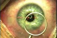
NEW INTRACOR PRESBYOPIA TREATMENT!!!!
THE MOST SIGNIFICANT INNOVATION OF THE RECENT 20 YEARS IN OPHTHALMOLOGY
THE TREATMENT OF AGE-RELATED VISUAL IMPAIRMENT HAS BEEN FOUND!
The treatment of "INTRACOR Presbyopia" which had been studied for years by the International Ophthalmology world having a high ratio of success in the treatment of age-related farsightedness impairment (presbyopia), emerging after the age of 40, was developed in Colombia by a team involving a Turkish doctor and was considered as the most important invention of the last two decades.
This method that had been studied for a long time was developed by the Head of Ophthalmology Department, Op. Dr. Sinan Göker, in Istanbul Cerrahi Hospital and the famous ophthalmologist Dr. Luis Ruiz Bogata who is amongst the doctors who had developed the Lasik Method for the purposes of myopia, astigmatism and hypermetropia.
Dr. Sinan Göker learned the Lasik technique Dr. Ruiz in 1997 that was used as an native method in eye operations and he was the first to apply it in Turkey.
There has been no regression seen or infection observed in the follow-ups performed following "INTRACOR Presbyopia Treatment" applied by Dr. Luis Ruiz and Op. Dr. Sinan Göker on more than 500 patients for approximately 6 months.
Un previous methods, this treatment, also called Intra COR, may be applied for both eyes. In contrast to the other examinations, it does not have an effect on distant sight.
No flap lysis, flap removing or tissue shaving is applicable. It heals up immediately (in 1-2 hours) as it is applied on the deep layers of the cornea.
The process takes 15-20 seconds following the narcotisation of the eyes with drops.
Post-treatment stinging or watering of eyes may be seen for 1-2 hours.
Following treatment, drops are used for 1-2 months. The patient may get to work on the following day.
Dry Eye
Dry eye patients have redness and watering of eyes as well as the feeling of sand in the eye. The most prominent characteristic is watering of eyes contrary to the term of "dry". There are 2 types of tears:
Basic Tear; It is continually and slowly secreted in to protect and wet the corneal surface. It is a balanced liquid consisting mucous, fatty, protein, enzyme, sucrose and antibodies.
Reflex Tear; Occurs in the cases of smoke, eye injuries, happiness and sadness.
Dry Eye Syndrome refers to the quality and quantity impairment of the basic tear.
Symptoms:
- Watering
- Itching-burning
- Blurred vision
- Redness
- Blurring
The reasons:
- Age-dependent
- Hormonal changes, especially postmenopausal
- Certain hypertension and allergy pills; antidepressants, sleeping tablets and analgesics.
- Air conditioner, smoke-filled ambients
- Reading, using computer, watching television
- Using contact lenses for a long period
Treatment:
Dry eye is hard to be treated completely once it develops. Generally, Synthetic tear drops, tear gels, Omega3 administration and lacrimal duct obstruction (Puntum plugs) are recommended as treatment. Cyclosporine preparations are used for extremely dry eyes.
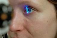
How do the doctors detect diopter values?
Nowadays, diopter values are automatically measured with apparatus called autorefractometer. However, these values are not assigned without testing with the patient. Devices called autophoropter are used for this process. The values are quickly detected by means of those devices. The measurements should be performed in two stages. First of all, measure the values without applying any drops into the eyes. Later on, apply a drop called cycloplegic which enlarges the eye pupil and relaxes the muscles around the lens, and wait for 40 minutes to ensure that it displays its effect. Thus, mismeasurement of values is prevented which could be a result of the accommodation of the lens.
What do the numbers in the prescription of glasses stand for?
Diopter values are expressed with a single value if there is no astigmatism available; as with 3 separate values if it is available. The first one is the spherical glass value, the second is the astigmatic glass value and the third one is the axis in which this astigmatic glass will be located. (+) stands for the convex glasses used for the improvement of hypermetropia; as (-) describes concave glasses used for myopia.
What is irregular astigmatism?
In case that the visual impairment originated the deformity in the cornea can be improved with spherocylindrical glasses, regular astigmatism is referred; as if it can not be improved irregular astigmatism is available. Irregular astigmatism is usually developed as a result of another disease (keratoconus, scar in the cornea, etc.). These patients may benefit using semi-hard gas-permeable contact lenses and their vision may get improved.
If you do not want to use two separate glasses for distant and near sight...
You will not have to shift glasses for distant and near sight due to the special glasses which we call progressive. However, those glasses are more expensive than the other ones and require a period of adaptation.
When I put my glasses on I feel dizziness as if I am stepping on a blank area.
Eyeglasses, especially with high diopter values and astigmatic glasses cause distortion in visions. While children and young people can easily get used to that vision the adaptation gets harder depending on the age. The period of adaptation varies between individuals. First of all, try to get used to glasses since they are the values with which you are able to see most sharply. If you can not get used to them, your doctor will reduce the values. However, it means a sum of sacrifice visual acuity. Certainly, all of them are acceptable in case there are not any mistakes done during assigning or making glasses. If you have any suspicions, you may have get your glasses measured by doctor.
What is amblyopia?
If visual acuity does not reach a certain level notwithstanding any particular disease in the eye and despite complete improvement with glasses, it means there is "amblyopia" in the eye. It occurs frequently depending on strabismus or the impairment of refraction difference between two eyes. If amblyopia is diagnosed and treated at early ages, vision level may get enhanced. Now, it is possible to succeed substantially at adult ages by means of Neurovision (treatment of amblyopia).